DJCAD MASTERS SHOW 2017: MSc Medical Art
The MSc Medical Art show demonstrates the use of a wide range of techniques, both traditional and digital, to educate and inform medical personnel and patients. In many cases highly complex ideas are demonstrated so clearly that even the non-medical viewer can understand the procedures and anatomy being explained.
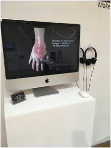
Inspired by his own experience undergoing chemotherapy, Ryan Berardi provides an on-screen visualisation to assist health care workers in alleviating the effects of one of the hazards of chemotherapy – extravasation, where the chemotherapy drug migrates dangerously to surrounding tissue and must be flushed out. Berardi’s lifelike animation explains the procedure clearly.
Claire Cooper’s work provides a digital teaching resource to enable medical students to understand the optic pathway, the complex means by which visual signals are passed from the eyes to the brain. The e-resource provides a 3D visualisation of the area which is clear enough for the non-medical viewer to grasp, plus a series of interactive questions and answers for those studying the topic at degree level. A similarly fascinating learning resource for medical students is provided by Elizabeth Paton’s interactive screen showing the effects of alcoholism on different organs. Using the computer mouse the diseased organs can be turned around on the screen for examination from all angles. Meanwhile a more traditional approach is seen in Amy Wedge’s detailed written analysis and physical models comparing the trauma caused by the impact of blunt or sharp force.
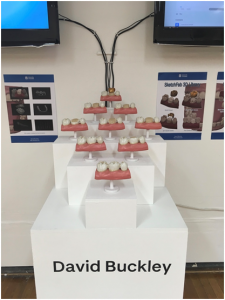
A number of exhibits form detailed teaching aids to assist in surgery. Stefanie Schutz employs augmented reality – using a HoloLens headset and interactive screen – to assist with ENT surgery. Again, interactive 3D modelling of body parts is shown on screen. Imi Ridley uses animation to demonstrate the complex procedure of endoscopic pituitary surgery. This view assists surgeons working in a small complex space using only an endoscope. Procedures with which the general public is perhaps more familiar are seen in David Buckley’s animation of three types of tooth crowning.
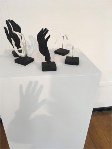
Two students focus on hand surgery in their projects. Natasha Mutch provides an on-screen 3D model of ligament reconstruction and tendon interposition, one of the most common hand surgeries. This resource can be used in doctor-patient consultations, enabling the latter to understand how the operation works and so alleviating anxiety. Thomas Parsons’ study evaluates the use of 3D printed models of the hand to enable the simulation of surgical procedures for finger joint replacement. His monochromatic 3D aids have the impact of delicate sculptural objects.
Catherine Truskolawski’s e-book documents the common injuries of the ankle and foot found in ballet dancers, while Arlene Yap offers a comic-book style website to educate and reassure parents and children about surgical procedures, especially anaesthesia.
The range of art techniques on offer, and the variety of medical issues that are illuminated in the exhibition, demonstrate the way in which artistic imagination combined with accurate vision can be of great benefit to students, practitioners and patients alike.


Leave a Reply